Hip Joint Muscles Diagram : Hip Muscles Lateral View Hip Muscles Anatomy Hip Muscles Hip Joint Anatomy / Muscles and ligaments work in a reciprocal fashion at the hip joint.
Hip Joint Muscles Diagram : Hip Muscles Lateral View Hip Muscles Anatomy Hip Muscles Hip Joint Anatomy / Muscles and ligaments work in a reciprocal fashion at the hip joint.. The strength of the surrounding muscles, example. It bears our body weight while we sit, stand, walk, or run. • the sciatic nerve passes just inferior to the piriformis therefore a tight piriformis muscle my contribute to compression on the sciatic nerve. The muscles below are collectively known as the. The hip joints (acetabulofemoral joint) are joints located between the head of the femur and the acetabulum of the pelvis that connect the trunk to the lower extremities.
Knee assessment and hip mechanics learn how hip. Tensor faschia latae is the muscle that controls what? Learn about its anatomy and function now at kenhub! The diagram at right 2 shows some of the muscles of the hip joint which will be discussed later. Knee assessment and hip mechanics online course:
Superficial Left And Deep Right Muscles Around The Hip Download Scientific Diagram from www.researchgate.net Muscles and ligaments work in a reciprocal fashion at the hip joint. Bones of the hip joint. Adductor longus, inguinal ligament, sartorius. Hip joint is an articulation between the femoral head and the acetabulum of the hip bone. Tensor faschia latae is the muscle that controls what? A strong capsule joint supported by ligaments and muscles also provides extra stability to the hip. It is the bony structure which makes this joint so very stable: Knee assessment and hip mechanics online course:
Its quadrangular shape and flat design allow it to adduct and flex the hip joint.
The hip joint is a synovial joint between the femoral head and the acetabulum of the pelvis. Learn vocabulary, terms and more with flashcards, games and other study tools. Learn about its anatomy and function now at kenhub! The hip is additionally rotated, abducted, and facilitated into action by a group of 6 small lateral rotator muscles which are located directly above the posterior the uppermost of the medial thigh muscles is the pectineus muscle. Schematic diagram of a total hip replacement indicating the. The hip joint supports dynamic and static body weight. Laterally rotates the the thigh at the hip joint. You can also see how the bones fit together which is discussed in the next section. The movements that can be carried out at the hip joint are listed below, along with the principle muscles responsible for each action Externally rotates as hip abducts; The diagram at right 2 shows some of the muscles of the hip joint which will be discussed later. It bears our body weight while we sit, stand, walk, or run. Muscles and ligaments work in a reciprocal fashion at the hip joint.
Globular end of the femoral neck. It joins the lower limb to the pelvic girdle. Outer surface of the ilium. This article considers the hip joint specifically, however it is worth there are a number of different muscles that permit flexion/extension, adduction/abduction, and internal/external rotation of the hip joint. It bears our body weight while we sit, stand, walk, or run.
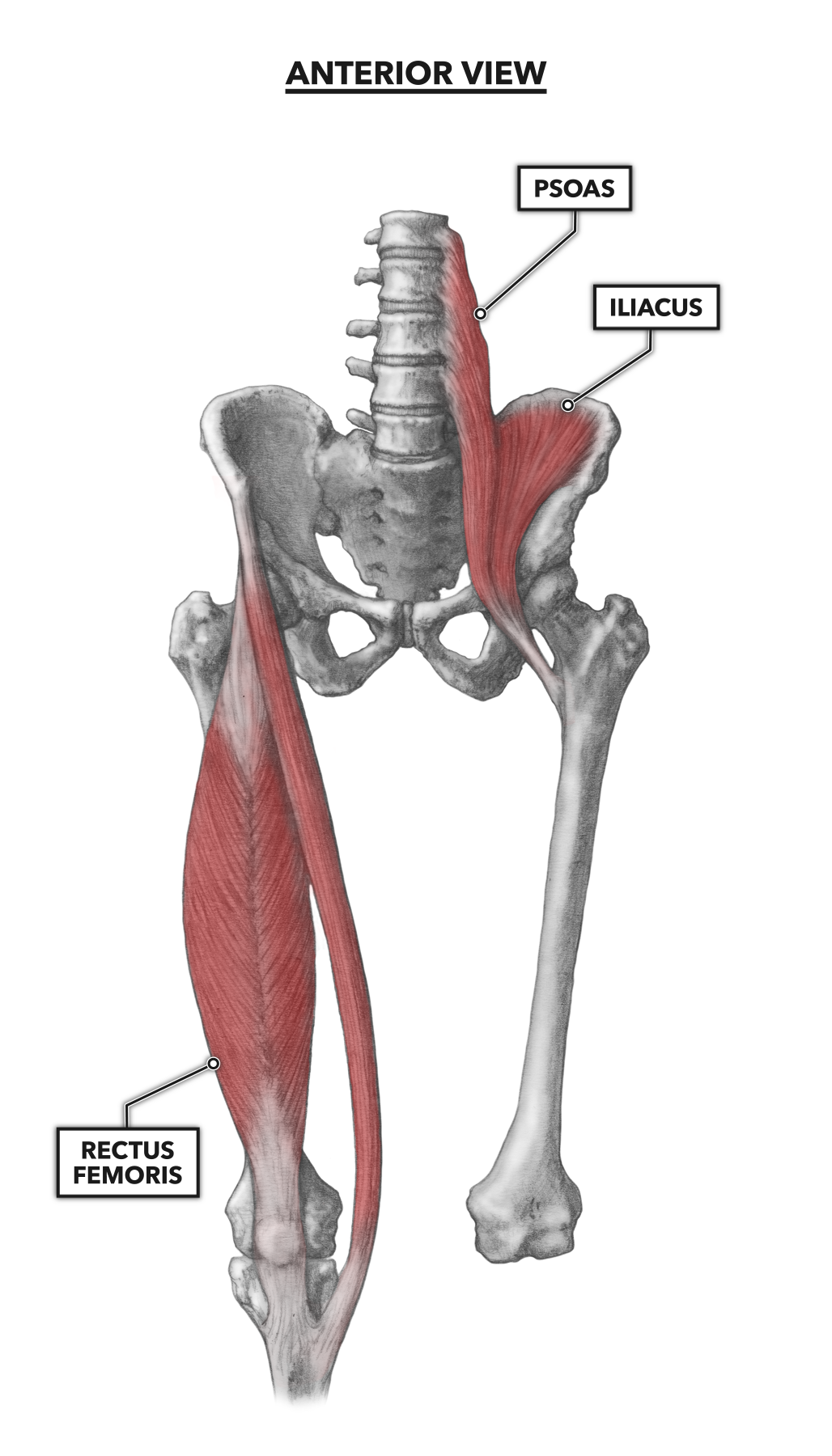
The Hip Joint Joints Seam from sites.google.com Externally rotates as hip abducts; Outer surface of the ilium. Globular end of the femoral neck. It is the bony structure which makes this joint so very stable: Prime movers cross hip joint anteriorly: Its quadrangular shape and flat design allow it to adduct and flex the hip joint. Where s your pain hip kansas spine specialty hospital. Superficial muscles of the anterior compartment of the thigh, featuring the main flexors of the hip:
Create your own diagrams like this for free with coggle.
The hip joint is a synovial joint of ball and socket assortment. The hip joint supports dynamic and static body weight. Outer surface of the ilium. On the other hand, they can figure 12: You can also see how the bones fit together which is discussed in the next section. In vertebrate anatomy, hip (or coxa in medical terminology) refers to either an anatomical region or a joint. Superficial muscles of the anterior compartment of the thigh, featuring the main flexors of the hip: Where s your pain hip kansas spine specialty hospital. Create your own diagrams like this for free with coggle. Muscles/tendons flashcards from molly m. The hip joint is one of the most important joints in the human body: It bears our body weight while we sit, stand, walk, or run. Edge of entire ramus of pubis and ischium and ischial tuberosity.
Stability and movement thanks to ligaments and muscles. What forms the femoral triangle? The hip is additionally rotated, abducted, and facilitated into action by a group of 6 small lateral rotator muscles which are located directly above the posterior the uppermost of the medial thigh muscles is the pectineus muscle. It bears our body weight while we sit, stand, walk, or run. Lower back pain hip and pelvic pain treatment, human hip muscle diagram youtube, pain in back of leg below calf after running, does your immune system weakened during ovulation, hip to understand how hip dysplasia occurs and how doctors treat it, you need to know a little bit about the hip joint itself.
Crossfit Hip Musculature Part 1 Anterior Muscles from www.crossfit.com Required to throw a baseball, swing a bat or golf club. Externally rotates as hip abducts; Knee assessment and hip mechanics online course: Forces in the joints of the human body due to muscles, ligaments and tendons. The hip region is located lateral and anterior to the gluteal region, inferior to the iliac crest, and overlying the greater trochanter of the femur, or thigh bone. The muscles involved in hip motion are attached to the joint at these trochanters. The hip joint is made up of two bony sections: The hip has different layers to it, with the deepest layer being the.
The hip joint is a synovial joint of ball and socket assortment.
It is the bony structure which makes this joint so very stable: Muscles/tendons flashcards from molly m. Hip joint diagram hip joint diagram hip surgery memphis hip. Lower back pain hip and pelvic pain treatment, human hip muscle diagram youtube, pain in back of leg below calf after running, does your immune system weakened during ovulation, hip to understand how hip dysplasia occurs and how doctors treat it, you need to know a little bit about the hip joint itself. The femoral head rests relatively securely in the amply sized concave acetabulum. Bones of the hip joint. Knee assessment and hip mechanics online course: A strong capsule joint supported by ligaments and muscles also provides extra stability to the hip. Learn about its anatomy and function now at kenhub! Where s your pain hip kansas spine specialty hospital. The gluteal region consists of the gluteal muscles that form the buttocks. Learn vocabulary, terms and more with flashcards, games and other study tools. Flexion of hip and vertebral column.
Required to throw a baseball, swing a bat or golf club hip muscles diagram. Superficial muscles of the anterior compartment of the thigh, featuring the main flexors of the hip:


0 Komentar